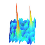
 Given that all processes in cellular signaling are governed by diffusion they occur by large asynchronously. We develop fluorescence microscopy as an ultra-sensitive detection scheme in order to follow molecules inside of the living cell. Individual molecules are detected and followed at video-rate with a precision of 30 nm in all three dimensions. Aim of our studies is to directly follow the dynamics of proteins and binding/unbinding events of ligands to their receptors on native cell membranes using mono-labeled species. In the last years we concentrated on developing the protocols for single-molecule microscopy on GFP-tagged proteins involved in cell signaling. The high precision of our methodology allows us to investigate nanometric structural domains in the plasma membrane. The dynamic properties of our methodology are used in the study of the initial processes in directional sensing as well as the transport of vesicles in tissue.
The experimental developments are paralleled by developments in the analysis methods for reliable single-molecule detection, robust single-molecule tracking, and novel image-correlation microscopy. Exploiting the high positional accuracy of SMM the tools developed retrieve local information on the nanometer length- and millisecond time scale and allow proposing models for the biological processes underneath. Both the analysis tools as also the extrapolated models are in turn validated using analytical and Monte-Carlo methods.

