 The process by which cells detect a gradient of a chemoattractant and subsequently initiate movement towards the source is called chemotaxis. This complex cellular response involves several interrelated processes, including directional sensing, polarized cytoskeletal organization and motility. Chemotaxis is involved in processes like neurogenesis, angiogenesis and many morphogenic processes. Chemotaxis also plays an important role in neutrophils as they roam the human body in search for infections.
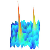
We use the slime mold Dictyostelium discodeum (Dicty) to study chemotaxis processes. The figure to the left shows Dicty cells migrating towards a cyclic adenosine mono-phosphate (cAMP) secreting micropipette (cAMP is a natural chemoattractant for Dictys). Upon stimulation by a cAMP gradient, which may be as small as a 2% across the cell body, the cells readily move towards the source of cAMP. Biochemical studies have shown that the intracellular organization of the cells engaged in chemotaxis is highly polar. Thus cells are able to translate a very shallow extracellular gradient into a steep, polarized intracellular response. The question we are trying to answer is: “How does this work?”
Using single-molecule microscopy (tracking of individual molecules as they diffuse through the cell membrane of life cells) we analyze the difference of the dynamic behavior of important proteins within the signaling pathway (cAMP receptor, G-proteins, small GTPases) between the cell’s anterior and posterior. We have shown that indeed the dynamical behavior of the receptor is already polarized and specific to G-protein coupling. From these findings we concluded that polarization of the dynamic behavior will play a central role in the signal amplification of the cAMP gradient.
A detailed analysis of the protein mobility (cAMP receptor, G-proteins, small GTPases) under various conditions and in various knockout background will reveal whether membrane nano-structuring is important for the biological function in this system. Our experimental findings are backed-up by finite-element modeling in order to gain a quantitative understanding of the reactions that ultimately lead to the chemotaxis response.
in collaboration with:
Ewa Snaar-Jagalska, Department of Cell Biology
latest results:
cAR internalization: Arnauld Serge, Sandra de Keijzer, Freek Van Hemert, Mark R. Hickman, Dale Hereld, Herman P. Spaink, Thomas Schmidt & B. Ewa Snaar-Jagalska. “Quantification of GPCR internalization by single-molecule microscopy in living cells.”, Integrative Biology (2011) 3:675
The cAR signalosome: Freek van Heemert, Milena Lazova, B.Ewa Snaar-Jagalska & Thomas Schmidt. “Mobility of G proteins is heterogeneous and polarized during chemotaxis.”, J. Cell Sci. (2010) 123:2922