Hemidesmosome mechanics, JCB 219, e201904137

Wang, W., A. Zuidema, L. te Molder, L. Nahidiazar, L. Hoekman, T. Schmidt, S. Coppola, and A. Sonnenberg. 2020. Hemidesmosomes modulate force generation via focal adhesions. The Journal of Cell Biology 219(2).
[DOI]

Abstract
Hemidesmosomes are specialized cell-matrix adhesion structures that are associated with the keratin cytoskeleton. Although the adhesion function of hemidesmosomes has been extensively studied, their role in mechanosignaling and transduction remains largely unexplored. Here, we show that keratinocytes lacking hemidesmosomal integrin α6β4 exhibit increased focal adhesion formation, cell spreading, and traction-force generation. Moreover, disruption of the interaction between α6β4 and intermediate filaments or laminin-332 results in similar phenotypical changes. We further demonstrate that integrin α6β4 regulates the activity of the mechanosensitive transcriptional regulator YAP through inhibition of Rho–ROCK–MLC– and FAK–PI3K–dependent signaling pathways. Additionally, increased tension caused by impaired hemidesmosome assembly leads to a redistribution of integrin αVβ5 from clathrin lattices to focal adhesions. Our results reveal a novel role for hemidesmosomes as regulators of cellular mechanical forces and establish the existence of a mechanical coupling between adhesion complexes.

Posted in Publications | Leave a comment

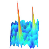
Brillouin Imaging: BOE 5, 2202 –

Coppola, S., T. Schmidt, G. Ruocco, and G. Antonacci. 2019. “Quantifying cellular forces and biomechanical properties by correlative micropillar traction force and Brillouin microscopy”. Biomed. Opt. Express 10(5):2202-2212.
[DOI] [pdf]

Cells sense and respond to external physical forces and substrate rigidity by regulating their cell shape, internal cytoskeletal tension, and stiffness. Here we show that the combination of micropillar traction force and noncontact Brillouin microscopy provides access to cell-generated forces and intracellular mechanical properties at optical resolution. Actin-rich cytoplasmic domains of 3T3 fibroblasts showed significantly higher Brillouin shifts, indicating a potential increase in stiffness when adhering on fibronectin-coated glass compared to soft PDMS micropillars. Our findings demonstrate the complementarity of micropillar traction force and Brillouin microscopy to better understand the relation between cell force generation and the intracellular mechanical properties.

Posted in Publications | Leave a comment

First Person: Veer Keizer –

Interview with Veer on GR mobility in JCS.

Posted in Member, News | Leave a comment

GR mobility: JCS 132, 2019

Keizer VIP, Coppola S, Houtsmuller AB, Geverts B, van Royen ME, Schmidt T, Schaaf MJM. Repetitive switching between DNA-binding modes enables target finding by the glucocorticoid receptor. J Cell Sci. 2019 Feb 25;132(5). pii: jcs217455.
[DOI] [pdf]

Posted in Publications | Leave a comment

hERG and cytoskeleton in PDAC: Cancer 23, 2019

Manoli S, Coppola S, Duranti C, Lulli M, Magni L, Kuppalu N, Nielsen N, Schmidt T, Schwab A, Becchetti A, Arcangeli A. The Activity of Kv 11.1 Potassium Channel Modulates F-Actin Organization During Cell Migration of Pancreatic Ductal Adenocarcinoma Cells. Cancers. 2019 Jan 23;11(2). pii: E135.
[DOI] [pdf]

Posted in Publications | Leave a comment